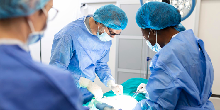
Coronary Angiography and Angioplasty
- Coronary Angiography and Angioplasty
- Dr. Rahul Katariya
Coronary angiography and angioplasty are medical procedures used to diagnose and treat heart conditions, particularly those related to the coronary arteries.
Coronary Angiography
Coronary angiography is a diagnostic procedure that uses X-ray imaging to see the heart's blood vessels. It is part of a general group of procedures known as cardiac catheterization.
Purpose:
To identify blockages or narrowing in the coronary arteries that could lead to chest pain (angina), heart attack, or other heart problems.
Procedure:
1. Preparation: The patient is usually awake but may be given a sedative. A local anesthetic is applied to numb the area where the catheter will be inserted.
2. Insertion: A thin, flexible tube called a catheter is inserted into an artery, typically in the groin or wrist.
3. Navigation: The catheter is carefully guided through the blood vessels to the coronary arteries.
4. Contrast Dye: A special dye (contrast medium) is injected through the catheter into the coronary arteries.
5. Imaging: X-ray images are taken as the dye flows through the arteries. These images help to highlight any blockages or narrowings.
Angioplasty
Angioplasty (also known as percutaneous coronary intervention or PCI) is a therapeutic procedure that involves opening up blocked or narrowed coronary arteries to restore proper blood flow to the heart muscle.
Purpose:
■ To treat coronary artery disease (CAD) and reduce symptoms such as chest pain and shortness of breath.
■ To minimize damage during or after a heart attack by restoring blood flow.
Procedure:
1. Preparation:Similar to angiography, a local anesthetic is applied, and the patient may receive a sedative.
2. Catheter Insertion: A catheter with a small balloon on its tip is inserted into an artery and guided to the narrowed part of the coronary artery.
3. Balloon Inflation: Once in place, the balloon is inflated to compress the plaque against the artery walls, widening the artery and restoring blood flow.
4. Stent Placement:In many cases, a stent (a small, wire mesh tube) is placed in the artery. The stent is expanded by the balloon and left in place to keep the artery open after the balloon is deflated and removed.
5. Completion:The catheter is removed, and the insertion site is closed and bandaged.
Both procedures are minimally invasive and are crucial in diagnosing and managing heart disease, potentially saving lives by preventing heart attacks and improving heart function.
